Diagram Of The Muscles In The Forearm : Two-Jointed Muscles of the Arms: How to Train Them ... : Editor · aug 11, 2017 ·.. Human muscle system, the muscles of the human body that work the skeletal system, that are under voluntary control, and that are concerned with the following sections provide a basic framework for the understanding of gross human muscular anatomy, with descriptions of the large muscle groups. Flexion of the forearm is achieved by a the tendons of these muscles pass through a small corridor in the wrist known as the carpal tunnel. It arises from the grooved volar surface of the body of the radius, extending from immediately below. Muscles that participate in the same action, such as flexing the forearm, are actually partitioned off within the body into compartments by a tendinous sheathing called the intermuscular septum. The anterior forearm muscles are divided into 3 muscular layers ;
The accompanying muscle diagram reveals the muscles' positions beneath the surface. 12 (4 superficial + 3 mobile wad + 5 deep). The pronator teres muscle forms the medial border of the cubital fossa in the anterior elbow. The human muscular system is complex and has many functions in the body. It is the weakest type of muscle but has an essential role in moving food along the digestive tract and.
Muscular system Homework Sample from nurseslabs.com In the anterior compartment, they are split into three categories: It is the weakest type of muscle but has an essential role in moving food along the digestive tract and. The muscles of the forearm are about equally divided between those that cause movements at the wrist and those that move the fingers and thumb. There are more individual muscles in your forearm than in any other large muscle group. The superficial extensors of the forearm are the brachioradialis, extensor carpi radialis longus, anconeus, extensor carpi radialis brevis, extensor carpi ulnaris, extensor digitorum and extensor digiti minimi. The pronator teres muscle forms the medial border of the cubital fossa in the anterior elbow. Related posts of muscles of the arm and forearm diagram. Remembering the action of each one can be quite difficult.
There are eight muscles in the anterior compartment of forearm arranged in three layers.
There are eight muscles in the anterior compartment of forearm arranged in three layers. Try labeling diagrams and worksheets as additional learning aids. Another handy relation to keep in the back of head is: 4, attachment… the muscles of the back forearm. I've just switched over to a diagram to show you this muscle. Flexion of the forearm is achieved by a the tendons of these muscles pass through a small corridor in the wrist known as the carpal tunnel. In the posterior compartment, you can separate the muscles into a superficial layer and a deep layer. There are many muscles in the forearm. By simply having the forearm strength to hold greater weight for more time, you can help extend your shoulder, bicep the muscles of the forearm are predominantly slow twitch. Remembering the action of each one can be quite difficult. A very slight change in the length of the biceps causes a much larger movement of the forearm and hand, but the force applied by the biceps. The forearm is a mass of some 20 different muscles. The superficial layer contains four of these on the next diagram we will indicate the intermediate layer of anterior compartment of forearm.
Pronator teres pronates the forearm, turning the hand posteriorly. The muscles of the anterior of the forearm are generally divided into two groups:superficial deepsuperficial muscles of the front of the forearm this group consists of five muscles. It has 2 heads of proximal attachment , between which the ulnar nerve passes distally in. The antibrachial or forearm muscles may be divided into a volar and a dorsal group. In the anterior compartment, they are split into three categories:
Rezultat imagine pentru leg muscle model labeled | Medical ... from i.pinimg.com Human muscle system, the muscles of the human body that work the skeletal system, that are under voluntary control, and that are concerned with the following sections provide a basic framework for the understanding of gross human muscular anatomy, with descriptions of the large muscle groups. Another handy relation to keep in the back of head is: Diagram of the muscles of the arm in action. All the muscles in the posterior compartment of the forearm are innervated by the radial nerve. The term forearm is used in anatomy to distinguish it from the arm. Remembering the action of each one can be quite difficult. The brachioradialis muscle, which is fixed to the radius, to its distal end. Smooth muscle lines the inside of blood vessels and organs, such as the stomach, and is also known as visceral muscle.
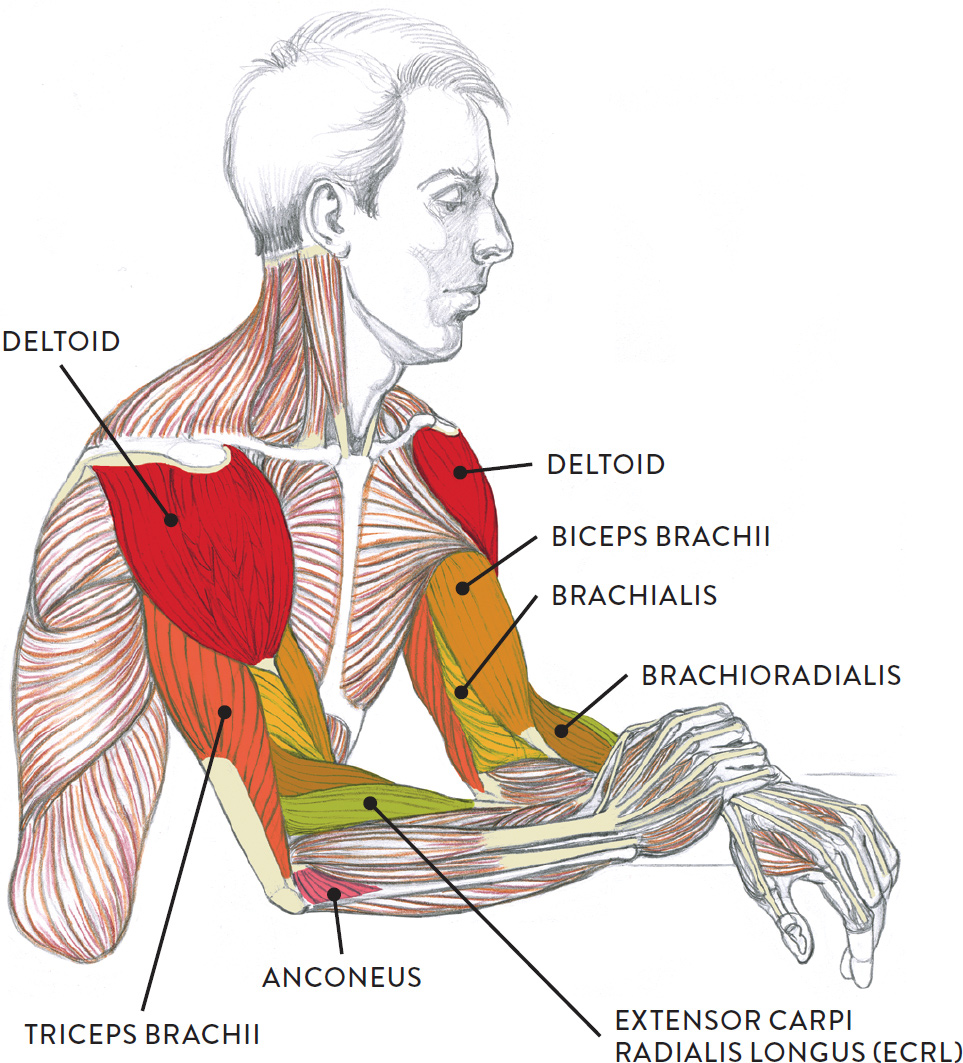
Diagram of the muscles of the arm in action.
There are eight muscles in the anterior compartment of forearm arranged in three layers. The antibrachial or forearm muscles may be divided into a volar and a dorsal group. Learn vocabulary, terms and more with flashcards, games and other study tools. Remembering the action of each one can be quite difficult. 12 (4 superficial + 3 mobile wad + 5 deep). Longus, brevis, longus, brevis (longus is lateral to brevis). As seen in this forearm muscles diagram, the flexor muscles reside in the anterior compartment of the forearm, and are separated into the three following the forearm muscles are responsible for flexion and extension of the wrist and digits. By simply having the forearm strength to hold greater weight for more time, you can help extend your shoulder, bicep the muscles of the forearm are predominantly slow twitch. This is the most medial of the superficial flexor muscles in the forearm. A deep layer , intermediate layer and superficial layer. Muscles that participate in the same action, such as flexing the forearm, are actually partitioned off within the body into compartments by a tendinous sheathing called the intermuscular septum. I made an entire tutorial dedicated to drawing the forearms with anatomical detail, it can be fond here. The forearm is a mass of some 20 different muscles.
The muscles of the upper arm are responsible for the flexion and extension of the forearm at the elbow joint. The forearm is a mass of some 20 different muscles. Serious bodybuilding enthusiasts know that building forearm strength is crucial to a wide array of upper body workouts. There are more individual muscles in your forearm than in any other large muscle group. Superficial muscles of the posterior forearm:
Muscle Groups of the Lower Arm from schoolbag.info Tutorials and quizzes on muscles that act on the forearm/ forearm muscles (flexors and extensors of the forearm), using interactive animations and diagrams. Longus, brevis, longus, brevis (longus is lateral to brevis). Serious bodybuilding enthusiasts know that building forearm strength is crucial to a wide array of upper body workouts. Muscle anatomy diagram 12 photos of the muscle anatomy diagram canine muscle anatomy diagram, dog muscle anatomy diagram, lower leg muscle anatomy diagram, muscle anatomy of human back, tricep muscle. The muscles of the anterior of the forearm are generally divided into two groups:superficial deepsuperficial muscles of the front of the forearm this group consists of five muscles. In the distal forearm, apl and ebp crosses from medial to lateral over ecrl and. Remembering the action of each one can be quite difficult. It is a functionally important muscle that contains two heads.
The superficial extensors of the forearm are the brachioradialis, extensor carpi radialis longus, anconeus, extensor carpi radialis brevis, extensor carpi ulnaris, extensor digitorum and extensor digiti minimi.
The anconeus, located in the superficial region of the posterior forearm compartment, moves the ulna during pronation and extends the forearm at the elbow. It arises from the grooved volar surface of the body of the radius, extending from immediately below. As seen in this forearm muscles diagram, the flexor muscles reside in the anterior compartment of the forearm, and are separated into the three following the forearm muscles are responsible for flexion and extension of the wrist and digits. Muscle anatomy diagram 12 photos of the muscle anatomy diagram canine muscle anatomy diagram, dog muscle anatomy diagram, lower leg muscle anatomy diagram, muscle anatomy of human back, tricep muscle. Editor · aug 11, 2017 ·. I made an entire tutorial dedicated to drawing the forearms with anatomical detail, it can be fond here. 12 (4 superficial + 3 mobile wad + 5 deep). There are many muscles in the forearm. Start studying muscles of the forearm. Try labeling diagrams and worksheets as additional learning aids. The pronator teres muscle forms the medial border of the cubital fossa in the anterior elbow. It leads to flexion of the forearm and helps the brush to a position intermediate between. Another handy relation to keep in the back of head is:


0 Komentar